Electron Microscopy of Beam-Sensitive Materials (B3)

Electron microscopy is used in Thrust B3 to study structural and local functional properties of materials in novel 3D systems manufactured within the Cluster. The properties of these materials have to be understood across several length scales – from the atomic structure and molecular packing, via phase morphology of fabricated heterogeneous systems, to the interaction of materials with bioorganic molecules, cells, and tissue.
Electron microscopy – in combination with energy dispersive X-ray spectroscopy, electron-energy-loss spectroscopy and cathodo-luminescence – provides high-resolution structural information as well as information on the local chemical composition, electronic and optical properties. Surface properties are assessed by scanning electron microscopy (SEM), while the study of internal material properties requires thin electron-transparent specimens, which are studied by (scanning) transmission electron microscopy ((S)TEM).
A tremendous variety of operating modes exists within these three classes of electron microscopies which will be adapted and optimized to the needs of the materials of interest. The overarching goal of Thrust B3 is to develop advanced techniques for sample preparation and for minimizing damage of electron-beam sensitive materials such as polymers and biological materials.
Further aims connected to this goal concern the contrast enhancement in these weakly scattering materials and the reconstruction of the three-dimensional structure by electron tomography to understand and optimize fabrication processes.
Thrust B3 will receive a large variety of different materials and 3D systems from other Thrusts of the Cluster. Tasks range from atomic-scale resolution imaging for structures produced in Research Area A to large-scale three-dimensional reconstruction of biological material with high spatial resolution in C.
Prof. Dr. Rasmus R. Schröder
Heidelberg University
rasmus schroeder@bioquant.uni-heidelberg.de
Images
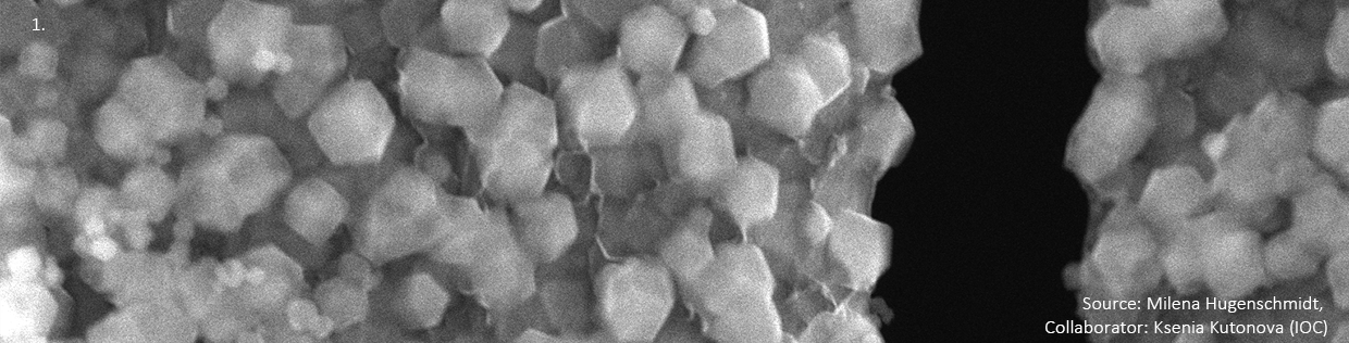
Description: Layer-by-layer-grown ZIF-8 (Source: Milena Hugenschmidt, Collaborator: Ksenia Kutonova (IOC))
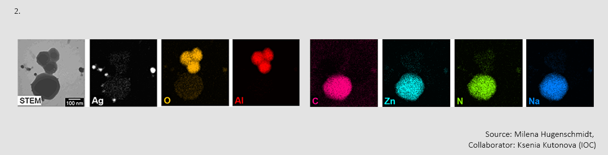
Description: EDX mapping of Ag/ZIF-8 (Source: Milena Hugenschmidt, Collaborator: Ksenia Kutonova (IOC))
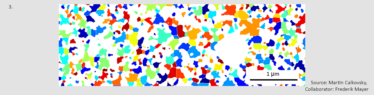
Description: Pore-size analysis of porous PETA structure via watershed algorithm (Source: Martin Calkovsky, Collaborator: Frederik Mayer)